The Normal Eye
It’s very easy to not appreciate your sight and to become complacent about being able to see, but each day in the UK, 100 people begin to lose their sight. RNIB has revealed that many harmful yet easily diagnosed eye problems still go undetected – a simple eye examination can reveal the truth.
It is vital that your eye examination is part of your overall health care. It will not only indicate the health of your eyes, but can also allow your Optometrist to detect early symptoms of general health problems, such as diabetes or high blood pressure.
The Normal Eye
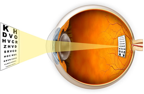
Emmetropia is the scientific name for normal vision. The light from an object focuses directly onto your retina forming a clear visual image. An individual with emmetropic vision can see distant and close objects clearly.
How we see
Seeing is similar to the process of taking pictures on a film with a camera which you then get developed. The retina is like a camera film which stores an image of what you are looking at. The image directed onto the retina is then sent to the brain where it is processed (like developing a camera film). Therefore you “see” in your brain with the light information sent to it from your eyes. Of course this process happens very very quickly.
How the eye works
You need light to see what is around you and to see colour. Light bounces off the objects you look at. These reflect different amounts of light which you see as different colours.
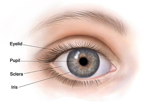
Front of the eye
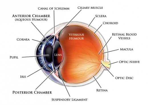
Light rays enter the front of your eye through the clear cornea and lens. It is very important that both the cornea and lens are clear as this allows the light to pass directly through the front of the eye to the retina. The cornea and lens bend light so that it can focus on the retina at the back of our eye and this gives you a clear, precise image. The cornea focuses the light towards your retina and the lens fine tunes the focussing of this light. Your tears form a protective layer at the front of the eye and also help to direct the light coming into your eye.
Your iris, the coloured circle at the front of our eye, changes the size of the pupil which allows different amounts of light into your eye. The pupil is the dark hole in the middle of the coloured part of your eye. The pupil gets smaller in bright conditions (it lets less light in) and bigger in dark conditions (it lets more light in).
Middle of the eye
The middle of your eye is filled with a jelly-like substance called the vitreous. The vitreous is clear and allows light to pass directly from the front to the back of our eye.
Back of the eye
The retina at the back of your eye is a light-sensitive layer which consists of rod and cone cells. These cells collect the light signals directed onto them and send them as electrical signals to the optic nerve at the back of our eye. Rod cells are concentrated around the edge of the retina. They help you to see things that aren’t directly in front of you, giving you a rough idea of what is around you. Rod cells help with your mobility and getting around by stopping you from bumping into things and they also enable you to see things in dim light and to see movement.
Cone cells are concentrated in the centre of your retina where the light is focused by the cornea and lens. This area is called the macula. Cone cells give you your detailed vision which is used when reading, watching TV, sewing and looking at people’s faces. They are also responsible for most of your colour vision. The optic nerve is made up of thousands of nerve fibres. These fibres pass the electrical signals along to your brain where they are processed into the image you look at. This connection is known as the visual system and develops through use from birth to around 7 years of age, after which it does not develop further.
Whilst the eye is natures most perfect invention when working properly, there are a number of conditions that can effect your vision if not discovered in time. To ensure our patients know the vital signs of these conditions we’ve compiled a guide to walk you through the signs and symptoms. Please click on the button below to find our more.